Endoscopy & Gastroscopy
Used to view and evaluate areas the horse including the upper respiratory tract (pharynx, larynx, guttural pouches and trachea), parts of the gastrointestinal tract (oesophagus, stomach, duodenum, rectum and small colon), urinary tract (bladder and urethra) and reproductive tract of mares (vagina, cervix and uterus).
This supports our work diagnosing horses displaying poor performance, abnormal respiratory function, upper airway obstructions, lung disease, gastric (stomach) ulcers and many urinary tract problems. Endoscopes are linked to a screen allowing live images and the ability for images to be recorded.
Gastric Ulcers are common and can effect horses of all types. The effects are believed to be associated with change in character, weight loss, changes in eating behaviour, poor performance and colic, although in many equines there are few or no apparent ill-effects.
The reliable way to diagnose gastric ulceration in horses is by gastroscopy (endoscopic examination). Gastroscopy supports the examination of the stomach and ulceration can be graded considering how deep widespread.
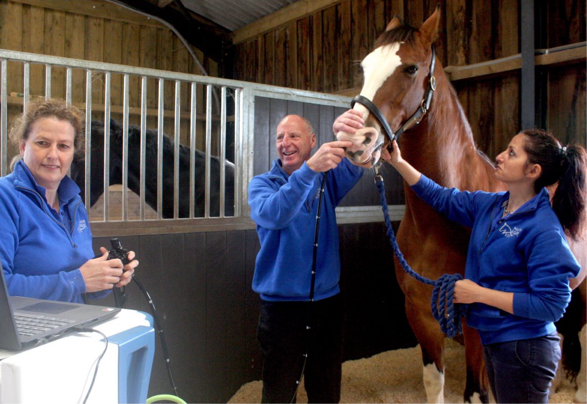
Gastroscopy – the procedure:
Performed under sedation, the gastroscope (a long flexible fibre optic camera) is passed up one of the horse’s nostrils, the horse then swallowing the gastroscope to pass into the stomach. Once inside the stomach, the stomach is inflated with air to allow full examination and the scope is manoeuvred to examine different regions of the stomach.
The horse or pony must be starved overnight before the gastroscopy, in order to empty the stomach of all food to allow good visualisation of all the mucosal surfaces.
The procedure is usually well-tolerated by the horse and takes around 20 minutes. Horses are ready to travel home when the sedation has worn off.
Airway Endoscopy – the procedure:
The endoscope is passed via a nostril enabling visual assessment of the nasal cavities. In each side of a horse’s nose there are 3 separate channels, within are complex folded bony structures – nasal conchae and ethmoids. The endoscope supports examination of these areas for a variety of abnormalities, from infection to cancerous growths or foreign bodies.
Further into the pharynx there are two openings into the eustachian tubes (connecting the back of the throat to the middle ear). In equines they are wide enough for an endoscope, they connect to the guttural pouches, which can develop infection or disease. One of the most common reasons to use an airway endoscopy is to look for signs of strangles infection in the guttural pouches.
In the pharynx we can examine the soft palate, epiglottis, pharyngeal lymph tissue, the larynx, arytenoids and vocal folds. Structures which are important for the normal function of a horse’s airway. Abnormality can be detrimental to normal air flow during exercise; serious for any horse that is a competing athlete. The most common abnormality is a partial paralysis of the horse’s larynx, causing a characteristic whistle noise when the horse breathes in during exercise.
Passing into the trachea, we can examine the quality and quantity of fluid exudate, helpful when looking for signs of lung disease. A lavage (tracheal wash) can be obtained from the trachea for examination of cells (cytology) and identification of bacterial infection. It is possible to pass the endoscope right down to where the trachea splits into the two large bronchi. A broncho-alveolar-lavage may also be used to wash cells from the lungs, including the lower bronchioles and alveolar air sacs. The wash fluid can then be analysed for accurate diagnosis of lung disease.